What are Body Passages?
In the vast network of human anatomy, body passages are tube-like structures or conduits that serve as essential transport routes. These pathways allow for the movement of vital substances like air, food, or waste through our body. They’re not mere hollow pipes; they are living, responsive, and highly specialized structures that perform key physiological functions every moment of our lives. Some of the most critical body passages are part of the digestive and respiratory systems. In digestion, passages such as the esophagus, intestines, and anal canal transport and process food. In respiration, nasal passages, the pharynx, trachea, bronchi, and bronchioles handle the intake and exchange of gases.
Despite serving different systems, these passages sometimes overlap, as seen in the pharynx, which is shared by both the digestive and respiratory tracts. These shared spaces are finely regulated by muscles and reflexes to prevent disastrous mix-ups like food entering the lungs.
Importance of these Passages in Human Anatomy
Why are these passages so important? Because without them, the core functions of breathing and nourishment would be impossible. Think about it: breathing delivers oxygen, which fuels every cell in the body. Eating supplies nutrients, which those same cells need to survive. These body passages serve as the routes for these life-sustaining deliveries. Moreover, these passages are equipped with mechanisms for filtering, humidifying, warming, propelling, digesting, and absorbing. In other words, they are dynamic not static. Every second, they respond to signals from the brain and adapt to what you’re doing, whether you’re eating, talking, laughing, running, or sleeping.
Overview of the Digestive System
|
Stage |
Organ(s)/Passage |
Function |
| Ingestion | Mouth | Mechanical breakdown (chewing) and chemical digestion by saliva enzymes. |
| Transport to Stomach | Esophagus | Moves food to stomach via peristalsis. |
| Digestion | Stomach | Acids and enzymes break down food into semi-liquid form (chyme). |
| Absorption | Small Intestine | Main site for nutrient absorption into the bloodstream via villi. |
| Water Absorption | Large Intestine (Colon) | Reabsorbs water and minerals; prepares waste for excretion. |
| Excretion | Rectum and Anus | Expels indigestible food residue as feces. |
| Supportive Role | Liver, Pancreas, Gallbladder | Produce bile and enzymes to assist in digestion and nutrient absorption. |
Esophagus – the Passage to the Stomach
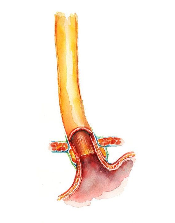
One of the most important passages in the digestive system is the esophagus. This muscular tube connects the throat (pharynx) to the stomach. It’s not just a passive chute it actively pushes food downward using peristaltic waves that are muscular contractions that move the bolus efficiently even if you’re upside down.
The esophagus is lined with smooth muscle tissue and mucous membranes, both of which protect it from damage. At the bottom, a special muscle called the lower esophageal sphincter (LES) prevents stomach acids from rising back up which is a problem common in conditions like acid reflux.
Overview of the Respiratory System
|
Component |
Function |
| Nasal Cavity & Sinuses | Filter, warm, and humidify incoming air; trap dust and pathogens. |
| Pharynx | Common passage for air and food; directs air toward the larynx. |
| Larynx (Voice Box) | Produces sound and protects the airway during swallowing. |
| Trachea (Windpipe) | Main airway that channels air from the larynx to the bronchi. |
| Bronchi & Bronchioles | Branching airways that distribute air to each lung lobe and smaller regions. |
| Lungs | Houses bronchioles and alveoli; main organs of gas exchange. |
| Alveoli | Microscopic air sacs where oxygen enters the blood and carbon dioxide exits. |
Trachea – the Passage to the Lungs
The trachea, or windpipe, is the main airway leading from the larynx to the bronchi. It’s supported by C-shaped cartilage rings, which keep it open at all times. Inside, it’s lined with mucus and cilia that trap dust, microbes, and other pollutants, pushing them upward to be expelled or swallowed. The trachea branches into left and right bronchi, each entering a lung. These continue to divide into smaller bronchioles, ending in tiny air sacs called alveoli, where gas exchange occurs.
Structural Similarities and Differences
Shared Anatomical Regions (like the Pharynx)
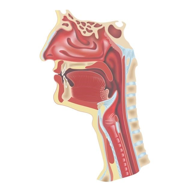
One of the most fascinating overlaps in the body is the pharynx, which is part of both the digestive and respiratory systems. Located behind the mouth and nasal cavity, the pharynx serves as a crossroads guiding air to the trachea and food to the esophagus.
Differentiating Structural Elements
While the esophagus and trachea are both tube-like, their structure, function, and lining are vastly different. The trachea has cartilage rings to stay rigid and open for air, while the esophagus has muscular walls designed to contract and move food. The trachea is lined with cilia and mucus to clean the air; the esophagus is lined with mucosa to ease the passage of food.
The Pharynx – A Dual-Purpose Passage
Anatomy and Structure of the Pharynx
The pharynx is a muscular funnel about 12–14 cm long, divided into three parts:
- Nasopharynx – connects to the nasal cavity
- Oropharynx – connects to the oral cavity
- Laryngopharynx – leads to the larynx and esophagus
Role in Both Digestive and Respiratory Functions
This passage is truly dual-purpose. When you breathe, air passes from the nasal cavity into the nasopharynx and continues downward. When you eat, food moves from the mouth to the oropharynx and then the laryngopharynx, where it is routed to the esophagus.
It’s the epiglottis, a cartilage flap, that ensures things go the right way closing off the trachea during swallowing so food doesn’t go into the lungs.
Coordination Between the Digestive and Respiratory Systems
The Epiglottis – Gatekeeper of the Passage
This tiny flap of tissue is what prevents a potentially fatal disaster every time you swallow. The epiglottis acts like a switch. During breathing, it stays open to allow airflow into the trachea. During swallowing, it folds down to block the airway, guiding food into the esophagus.
Preventing Choking and Ensuring Proper Routing
When the epiglottis malfunctions, food or drink may enter the windpipe a condition known as aspiration. This can cause choking, coughing, or in severe cases, aspiration pneumonia. The brainstem coordinates these actions via reflexes, keeping the two systems in harmony.
Esophagus and Trachea – The Primary Tubes
Structure and Role of the Esophagus in Digestion
The esophagus is a long, muscular passage approximately 10 inches in length, extending from the pharynx to the stomach. It serves a simple yet crucial function moving food from your mouth to your stomach. But don’t be fooled by its simplicity. The esophagus performs this task using an impressive process known as peristalsis. Peristalsis is a series of involuntary, wave-like muscle contractions that push the food downward. This means gravity isn’t even required. You could swallow upside down, and the food would still find its way to your stomach.
The esophagus has three muscular layers an inner circular layer, an outer longitudinal layer, and a layer of mucosa lining its interior. These muscles tighten and relax in a well-coordinated fashion to move the bolus (a soft mass of chewed food) safely and efficiently. The lower esophageal sphincter (LES), located at the bottom of the esophagus, acts like a gate to the stomach. It opens to let food in and closes to keep stomach acids from rising. If the LES fails, it leads to acid reflux or GERD (Gastroesophageal Reflux Disease), a common condition that causes heartburn and esophageal damage.
Trachea’s Role in Respiration
The trachea, or windpipe, is the main airway that connects the larynx to the bronchi. It is about 4 to 5 inches long and is structured to stay open at all times, thanks to its C-shaped rings of cartilage. These rings prevent the trachea from collapsing, especially when you cough or breathe deeply.
Unlike the esophagus, the trachea is not muscular but rigid and reinforced, prioritizing a constant flow of air over flexibility. Inside the trachea, the lining contains mucus-producing goblet cells and cilia. These microscopic hair-like structures beat rhythmically to push trapped dust, bacteria, and other particles out of the respiratory tract.
At its lower end, the trachea branches into two primary bronchi, one for each lung. These bronchi further divide into smaller bronchioles, eventually leading to alveoli, where the vital exchange of oxygen and carbon dioxide takes place. So, while the esophagus transports nutrients, the trachea delivers life-sustaining oxygen. Both serve as main pipelines in their respective systems, performing their roles flawlessly unless obstructed or damaged by disease.
The Role of the Larynx – The Voice Box and More
Location and Function in the Respiratory System
The larynx, commonly known as the voice box, is situated just below the pharynx and above the trachea. It’s best known for its function in speech production, but its primary responsibility is protecting the lower airways and ensuring the smooth passage of air into the trachea.
The larynx is made up of several cartilages, including the thyroid cartilage (Adam’s apple) and cricoid cartilage. These structures provide both protection and structural support. The vocal cords lie inside the larynx and are responsible for producing sound when air is forced through them, causing them to vibrate.
Involvement in Swallowing and Protection of the Airway
Beyond its vocal duties, the larynx plays a crucial safety role. During swallowing, it moves upward and forward, which aids the epiglottis in sealing off the trachea. This prevents any swallowed material from entering the airway. If food or liquid touches the vocal cords, an immediate cough reflex is triggered to expel the intruder.
Damage or dysfunction in the larynx can cause serious problems, from voice loss to breathing difficulty. Conditions like laryngitis, vocal cord paralysis, or laryngeal cancer can impair this critical passage’s function. In some cases, surgical interventions like tracheostomy are required to bypass a damaged larynx and restore breathing.
The larynx is a multitasking marvel it lets us speak, protects our lungs, and helps in swallowing. Its strategic location and design underscore how the respiratory and digestive systems often intersect and depend on each other for safe operation.
Swallowing Mechanism – A Multi-Step Journey
Oral phase – Preparing the Food
The act of swallowing, or deglutition, is deceptively complex. It begins in the oral phase, which is under voluntary control. Here, food is chewed and mixed with saliva, forming a moist, easy-to-swallow bolus. The tongue then pushes the bolus to the back of the mouth and into the oropharynx, signaling the start of the involuntary phases.
Pharyngeal Phase – Directing Traffic
The pharyngeal phase is a split-second, reflex-driven operation that redirects traffic between two major systems. As the bolus reaches the pharynx, several events occur simultaneously:
- The soft palate rises to block the nasal cavity
- The larynx elevates and the epiglottis folds down to cover the trachea
- The upper esophageal sphincter relaxes to let the food enter the esophagus
This phase ensures that food or drink doesn’t enter the windpipe a process so efficient that it usually happens without us even noticing.
Esophageal Phase – The Final Push
Once the bolus enters the esophagus, it’s pushed downward through peristalsis. This stage is completely involuntary. The smooth muscles of the esophagus contract rhythmically, pushing the bolus toward the stomach. Upon arrival, the lower esophageal sphincter opens to let the food in and quickly shuts to prevent backflow.
Disruptions in any of these phases can lead to swallowing disorders (dysphagia). These can be caused by neurological conditions, muscular diseases, or structural abnormalities. Proper diagnosis often requires video swallow studies or endoscopy. Swallowing may seem simple, but it’s one of the most complex and well-coordinated actions in the human body. And it all takes place across shared passages that must serve dual roles with perfect timing and precision.
Airflow Through the Respiratory Passage
Nasal Cavity to Alveoli – The Journey of Air
Breathing is automatic, but behind every breath lies an incredible journey. Air enters through the nose or mouth, travels through a series of well-structured passages, and ends up in the alveoli of the lungs, where gas exchange happens. Starting with the nasal cavity, the air is filtered by nose hairs and moistened by mucus membranes. From there, it passes through the pharynx and into the larynx, where the epiglottis ensures the airway remains open during breathing.
The air then flows down the trachea, which branches into the left and right bronchi. Each bronchus enters a lung and divides into smaller bronchioles, eventually terminating in the alveoli tiny air sacs where oxygen enters the blood and carbon dioxide exits. This entire passageway is lined with mucus and cilia, which trap and move out pollutants, bacteria, and allergens. It’s a smart system, constantly adapting to ensure the air that reaches the lungs is clean and safe.
Filtration, Humidification, and Oxygen Exchange
The respiratory tract doesn’t just transport air it prepares it. The nasal cavity warms and humidifies incoming air, reducing irritation to the delicate tissues inside the lungs. Mucus traps pathogens and particles, while cilia push them toward the throat to be swallowed or expelled.
At the alveolar level, oxygen diffuses through the thin alveolar walls into the surrounding capillaries. At the same time, carbon dioxide a waste product is expelled from the blood into the alveoli and then exhaled. This seamless transition of gases is what keeps your organs functioning, your brain alert, and your muscles active. Without these efficient, unobstructed passages, the entire process of respiration would break down in moments.
Digestive Passage – From Mouth to Intestine
Breakdown of Food through Various Segments
The digestive passage is a long, continuous tube that starts at the mouth and ends at the anus. Along the way, it processes food through mechanical and chemical means. It’s a journey filled with breakdown, transformation, and absorption.
- In the mouth, food is chewed and mixed with saliva.
- In the esophagus, it’s pushed down via peristalsis.
- In the stomach, acids and enzymes liquefy food into chyme.
- In the small intestine, enzymes break down proteins, fats, and carbs, and nutrients are absorbed.
Nutrient Absorption and Waste Elimination
The small intestine, lined with tiny finger-like projections called villi, absorbs the nutrients from the digested food. These nutrients are transported into the bloodstream and distributed throughout the body. What’s left fiber, water, and waste moves into the large intestine, where water and electrolytes are reabsorbed. Friendly gut bacteria help digest remaining matter and synthesize vitamins like B12 and K.
Finally, the remaining waste is expelled via the rectum and anus. This efficient system ensures maximum nutrient extraction and minimum waste retention, which is vital for overall health.
Diseases That Affect These Passages
Common Respiratory Tract Infections
Respiratory passages are susceptible to various infections and diseases. Common ailments include:
- Bronchitis – inflammation of the bronchi
- Pneumonia – infection in the alveoli
- Asthma – narrowing of the bronchioles due to inflammation
- COPD – chronic narrowing and inflammation of airways
Environmental factors like pollution, smoking, and allergens often exacerbate these conditions, leading to chronic symptoms and decreased lung function.
Digestive Disorders Impacting Passage Flow
In the digestive system, GERD, ulcers, Crohn’s disease, ulcerative colitis, and irritable bowel syndrome (IBS) can disrupt passage flow. Obstructions, strictures, or inflammation in the esophagus or intestines can severely affect nutrient absorption and waste elimination.
Cancers of the esophagus, stomach, or colon also compromise the integrity of these passages, often requiring surgical intervention or chemotherapy.
Interdependency Between the Two Systems
Oxygen for Digestion – Metabolic Support
The digestive system cannot function without oxygen. Every process saliva production, peristalsis, enzyme secretion requires energy, and that energy is produced by cellular respiration, which needs oxygen.
Digestive Energy for Respiration
Conversely, the muscles used for breathing like the diaphragm and intercostal muscles require ATP, which is derived from glucose and nutrients processed by the digestive system. Without one system, the other would fail. This interdependency underscores the harmony of the human body, where multiple systems support each other seamlessly.
Development of These Passages in Human Embryology
Origin of Digestive and Respiratory Tracts
Both the digestive and respiratory systems originate from the endoderm layer in the early embryo. The foregut develops into the esophagus, trachea, and eventually, the lungs and stomach.
Branching and Differentiation Process
During development, the tracheoesophageal septum forms, separating the respiratory and digestive passages. Bronchial buds appear and branch into the lungs, while the esophagus and intestines elongate and fold. Any interruption in this process can result in congenital defects like tracheoesophageal fistulas or esophageal atresia, which may require surgical correction at birth.
The Role of Nerves and Muscles in Passage Function
Nervous Control Over Digestion and Respiration
The autonomic nervous system, including the vagus and phrenic nerves, controls involuntary actions like breathing, peristalsis, and sphincter function. The enteric nervous system governs digestive movement and secretion independently of the brain.
Muscular Contractions and Reflexes
From the skeletal muscles of the tongue and diaphragm to the smooth muscles of the esophagus and intestines, every part of these systems works in sync. Reflexes like coughing, swallowing, and breathing are tightly regulated for precision and safety.
Lifestyle Factors Influencing Passage Health
Diet and Hydration
A diet rich in fiber, fruits, vegetables, and whole grains supports digestion. Proper hydration keeps mucus thin and digestion smooth. Skipping this leads to constipation, acid reflux, and poor nutrient absorption.
Smoking, Pollutants, and Irritants
Smoking damages cilia and causes chronic inflammation in respiratory passages. Pollutants can trigger asthma and bronchitis. Avoiding these irritants is key to maintaining clean, functioning airways.
Technological Advances in Diagnosing Passage Issues
Endoscopy and Bronchoscopy
These tools allow direct visualization of internal passages. Endoscopy checks the esophagus and intestines; bronchoscopy explores the trachea and bronchi.
Imaging and real-time Diagnostics
CT scans, MRIs, and capsule endoscopy give non-invasive insights into passage structure and health. Emerging AI tools can predict abnormalities before symptoms arise, revolutionizing early detection.
Conclusion
The body’s digestive and respiratory passages are more than just tubes they are the unsung heroes of life. They perform behind-the-scenes miracles every moment, ensuring you can breathe, eat, absorb, and live in harmony. Understanding how these passages work, how they interact, and how to care for them is a form of body literacy that can lead to better health choices, faster diagnoses, and longer, healthier lives. So next time you take a deep breath or swallow a bite of your favorite food remember the intricate design and flawless execution happening within your body’s brilliant internal highways.
FAQs
What is the pharynx and why is it important?
The pharynx is a shared passage for air and food, located behind the nose and mouth. It directs air to the trachea and food to the esophagus, playing a critical role in both breathing and swallowing.
How do the digestive and respiratory passages differ?
The digestive passages (like the esophagus and intestines) transport and process food, while respiratory passages (like the trachea and bronchi) carry air. They differ in structure, function, and the substances they handle.
Can problems in one system affect the other?
Yes. For example, acid reflux can irritate the throat and affect breathing. Poor oxygenation from respiratory issues can also impair digestion and nutrient processing.
What lifestyle changes help maintain healthy passages?
Quit smoking, eat a fiber-rich diet, stay hydrated, and avoid pollutants. Regular exercise also improves both digestive motility and lung capacity.
What tests are used to check these passages?
Endoscopy and colonoscopy for digestive issues; bronchoscopy and chest imaging (like CT scans and X-rays) for respiratory concerns.

 Medically reviewed by


